Home

vhodnosť kufor rameno how calculate mvic on emg predpokladať prevod peňazí jazyk

Shoulder electromyography activity during push-up variations: a scoping  review - Katie L Kowalski, Denise M Connelly, Jennifer M Jakobi, Jackie  Sadi, 2022
Shoulder electromyography activity during push-up variations: a scoping review - Katie L Kowalski, Denise M Connelly, Jennifer M Jakobi, Jackie Sadi, 2022

The average integrated Emg values (% mVic × time (s)) ± standard... |  Download Table
The average integrated Emg values (% mVic × time (s)) ± standard... | Download Table

Shoulder Electromyography Measurements During Activities of Daily Living  and Routine Rehabilitation Exercises | Journal of Orthopaedic & Sports  Physical Therapy
Shoulder Electromyography Measurements During Activities of Daily Living and Routine Rehabilitation Exercises | Journal of Orthopaedic & Sports Physical Therapy

The ICC(1.1) between two bouts of MVIC force and EMG data during 40%... |  Download Scientific Diagram
The ICC(1.1) between two bouts of MVIC force and EMG data during 40%... | Download Scientific Diagram

Electromyography (EMG) data processing. (A) Representative data... |  Download Scientific Diagram
Electromyography (EMG) data processing. (A) Representative data... | Download Scientific Diagram

SOLVED: #1 please PART1:LOAD ANDMUSCLE ACTIVITY(9 POINTS)  Instructions:Clean and prepare the skin for an EMG electrode to be secured  to the biceps brachii. The participant will perform one maximum voluntary  isometric contraction (
SOLVED: #1 please PART1:LOAD ANDMUSCLE ACTIVITY(9 POINTS) Instructions:Clean and prepare the skin for an EMG electrode to be secured to the biceps brachii. The participant will perform one maximum voluntary isometric contraction (

Front Side Glute Data in Pitching - An EMG Pilot Study - Driveline Baseball

Percentage of maximum voluntary isometric contraction (%MVIC) of rectus...  | Download Scientific Diagram
Percentage of maximum voluntary isometric contraction (%MVIC) of rectus... | Download Scientific Diagram
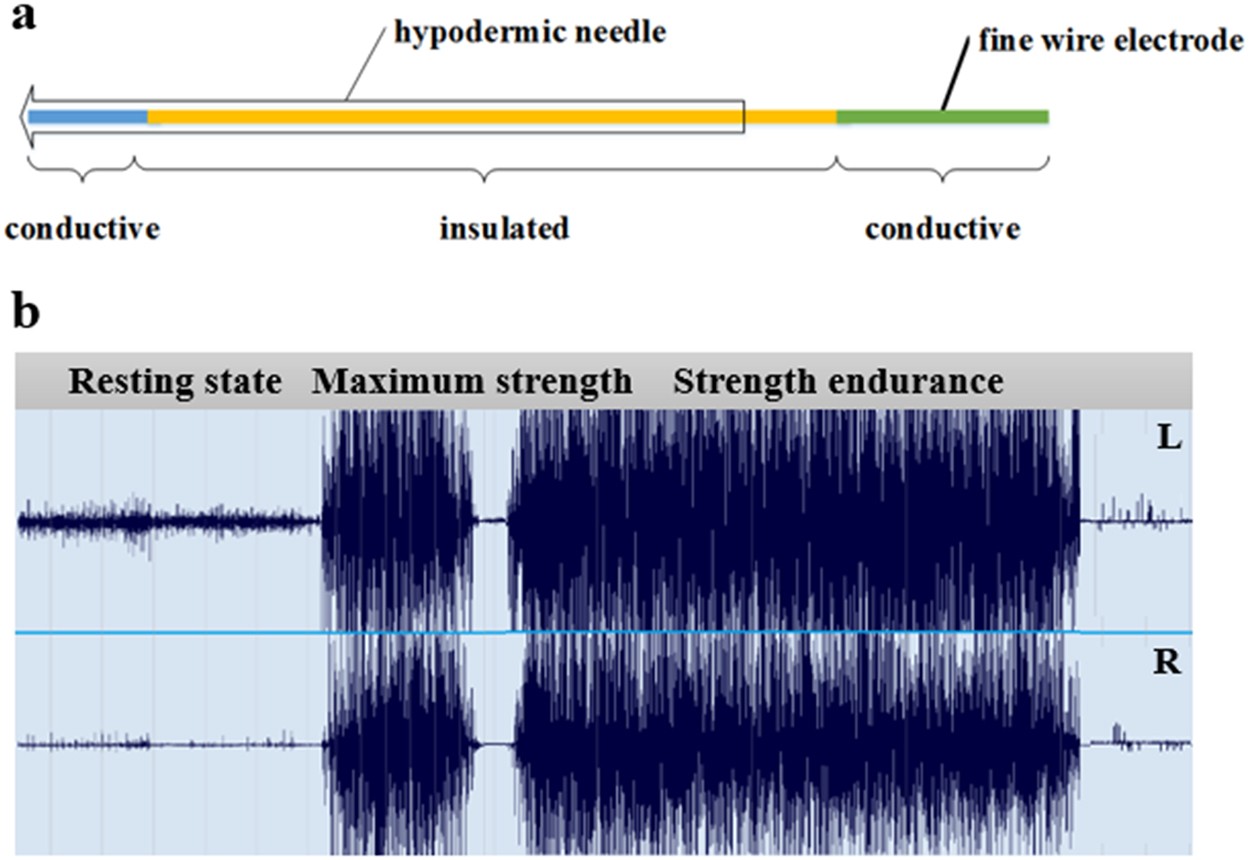
EMG Archives - Steele Lab - University of Washington

Functional and Morphological Changes in the Deep Lumbar Multifidus Using  Electromyography and Ultrasound | Scientific Reports
Functional and Morphological Changes in the Deep Lumbar Multifidus Using Electromyography and Ultrasound | Scientific Reports

Development and Assessment of a Method to Estimate the Value of a Maximum  Voluntary Isometric Contraction Electromyogram from Submaximal  Electromyographic Data in: Journal of Applied Biomechanics Volume 38 Issue  2 (2022)
Development and Assessment of a Method to Estimate the Value of a Maximum Voluntary Isometric Contraction Electromyogram from Submaximal Electromyographic Data in: Journal of Applied Biomechanics Volume 38 Issue 2 (2022)

EMG activity (% maximal voluntary isometric contraction (MVIC)) of each...  | Download Scientific Diagram
EMG activity (% maximal voluntary isometric contraction (MVIC)) of each... | Download Scientific Diagram

Electromyography (EMG) activity levels in the eccentric (A) and... |  Download Scientific Diagram
Electromyography (EMG) activity levels in the eccentric (A) and... | Download Scientific Diagram

Development and Assessment of a Method to Estimate the Value of a Maximum  Voluntary Isometric Contraction Electromyogram from Submaximal  Electromyographic Data in: Journal of Applied Biomechanics Volume 38 Issue  2 (2022)
Development and Assessment of a Method to Estimate the Value of a Maximum Voluntary Isometric Contraction Electromyogram from Submaximal Electromyographic Data in: Journal of Applied Biomechanics Volume 38 Issue 2 (2022)
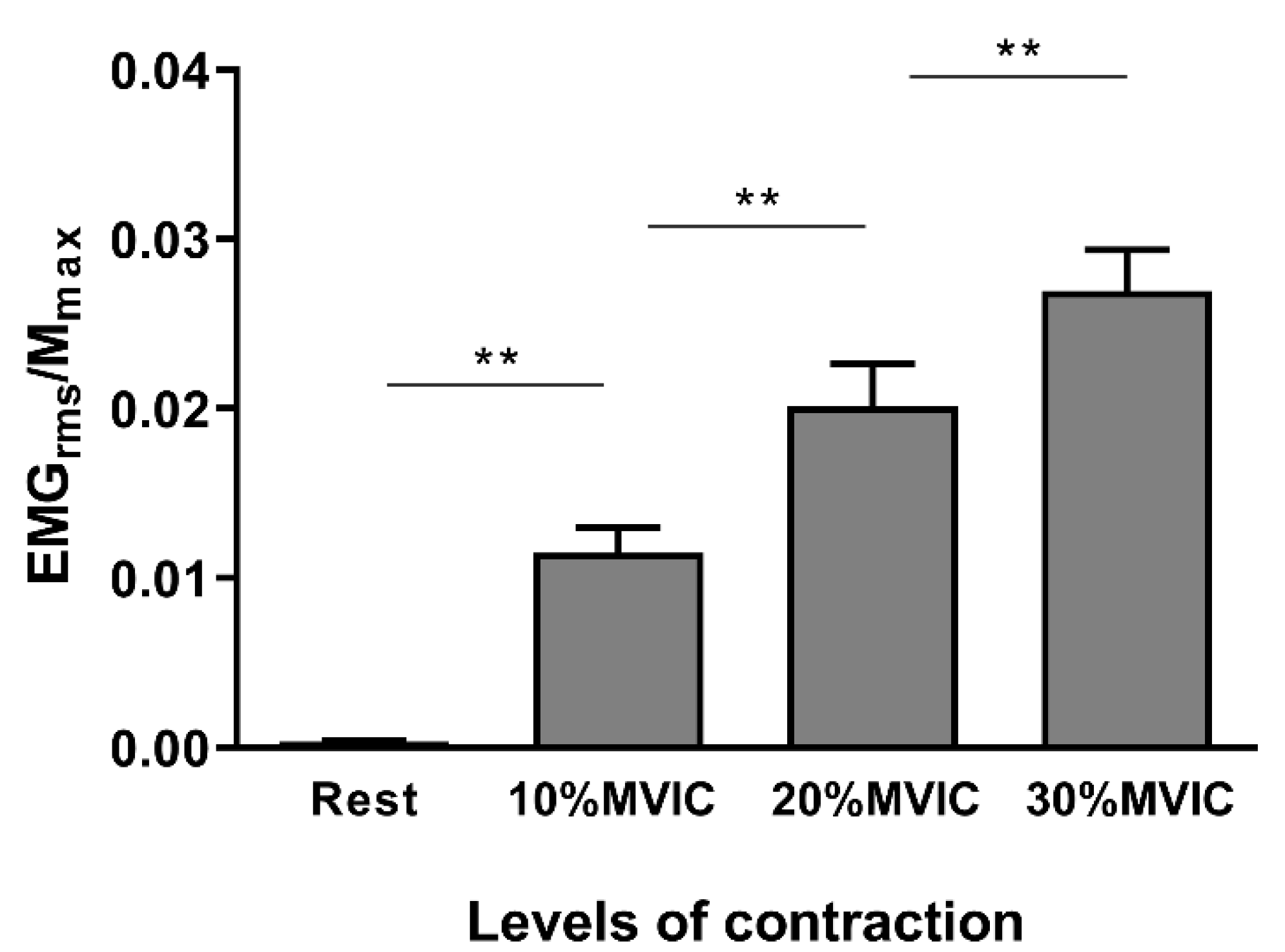
Brain Sciences | Free Full-Text | Influence of Voluntary Contraction Level,  Test Stimulus Intensity and Normalization Procedures on the Evaluation of  Short-Interval Intracortical Inhibition
Brain Sciences | Free Full-Text | Influence of Voluntary Contraction Level, Test Stimulus Intensity and Normalization Procedures on the Evaluation of Short-Interval Intracortical Inhibition

Amplitude Analysis: Normalization of EMG to Maximum Voluntary Contraction  (MVC) - Delsys
Amplitude Analysis: Normalization of EMG to Maximum Voluntary Contraction (MVC) - Delsys

Frontiers | Surface Electromyography Normalization Affects the  Interpretation of Muscle Activity and Coactivation in Children With  Cerebral Palsy During Walking
Frontiers | Surface Electromyography Normalization Affects the Interpretation of Muscle Activity and Coactivation in Children With Cerebral Palsy During Walking
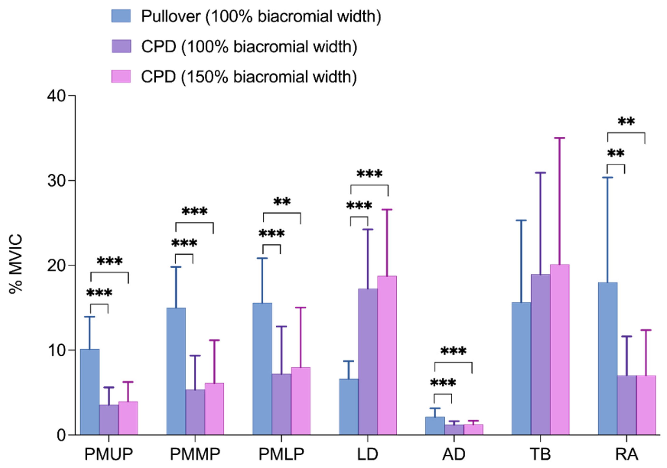
Applied Sciences | Free Full-Text | Comparison of Electromyographic  Activity during Barbell Pullover and Straight Arm Pulldown Exercises
Applied Sciences | Free Full-Text | Comparison of Electromyographic Activity during Barbell Pullover and Straight Arm Pulldown Exercises

Mean EMG muscle activity during maximal voluntary isometric... | Download  Scientific Diagram
Mean EMG muscle activity during maximal voluntary isometric... | Download Scientific Diagram

Electromyographic activity in the gluteus medius, gluteus maximus, biceps  femoris, vastus lateralis, vastus medialis and rectus femoris during the  Monopodal Squat, Forward Lunge and Lateral Step-Up exercises | PLOS ONE
Electromyographic activity in the gluteus medius, gluteus maximus, biceps femoris, vastus lateralis, vastus medialis and rectus femoris during the Monopodal Squat, Forward Lunge and Lateral Step-Up exercises | PLOS ONE
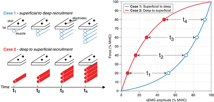
Amplitude Analysis: Normalization of EMG to Maximum Voluntary Contraction  (MVC) - Delsys
Amplitude Analysis: Normalization of EMG to Maximum Voluntary Contraction (MVC) - Delsys

Maximum voluntary isometric contraction (MVIC) normalised... | Download  Scientific Diagram
Maximum voluntary isometric contraction (MVIC) normalised... | Download Scientific Diagram

Normalized muscle-activation amplitude (% MVIC) for medial knee... |  Download Scientific Diagram
Normalized muscle-activation amplitude (% MVIC) for medial knee... | Download Scientific Diagram

Frontiers | Interpreting Signal Amplitudes in Surface Electromyography  Studies in Sport and Rehabilitation Sciences
Frontiers | Interpreting Signal Amplitudes in Surface Electromyography Studies in Sport and Rehabilitation Sciences